School of Engineering and Sciences
Department of Chemistry

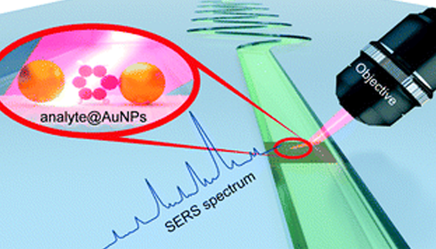
The Department of Chemistry is glad to announce that Dr J P Raja Pandiyan has published a paper titled ” Microfluidics and surface-enhanced Raman spectroscopy, a win-win combination?” in the journal ‘Lab on a Chip’ having an impact factor of 6.79 in collaboration with researchers from different universities across India, Germany, and Japan.
Abstract of the Research
With the continuous development in nanoscience and nanotechnology, analytical techniques like surface-enhanced Raman spectroscopy (SERS) render structural and chemical information of a variety of analyte molecules in ultra-low concentration. Although this technique is making significant progress in various fields, the reproducibility of SERS measurements and sensitivity towards small molecules are still daunting challenges. In this regard, microfluidic surface-enhanced Raman spectroscopy (MF-SERS) is well on its way to join the toolbox of analytical chemists. This review article explains how MF-SERS is becoming a powerful tool in analytical chemistry. We critically present the developments in SERS substrates for microfluidic devices and how these substrates in microfluidic channels can improve the SERS sensitivity, reproducibility, and detection limit. We then introduce the building materials for microfluidic platforms and their types such as droplet, centrifugal, and digital microfluidics. Finally, we enumerate some challenges and future directions in microfluidic SERS. Overall, this article showcases the potential and versatility of microfluidic SERS in overcoming the inherent issues in the SERS technique and also discusses the advantage of adding SERS to the arsenal of microfluidics.
About the Raman Research Group at SRM AP
Raman spectroscopy, invented by Sir CV Raman in 1928 and got Nobel Prize in 1930, is a vibrational spectroscopic technique that works based on the principle of inelastic scattering of light. Surface-Enhanced Raman spectroscopy (SERS) is one of the modern analytical techniques which can detect chemical and biomolecules in an ultra-low concentration. The research group is working on the development of the SERS technique to address the issues in food, environmental, energy and biological science.
The newly developed SERS substrates are mainly used for the detection of biological samples for disease diagnosis, food samples to ensure food safety, water samples to study the contamination and pollution rate. These studies can make meaningful social changes and improvements.
 Dr Nimai Mishra, Assistant Professor, Department of Chemistry, SRM University-AP, Andhra Pradesh, along with his research group comprising of students pursuing PhD under his supervision, Ms VG Vasavi Dutt, Mr Syed Akhil, Mr Rahul Singh, and Mr Manoj Paalabathuni have published a research article titled “High-Quality CsPbX3 (X = Cl, Br, or I) Perovskite Nanocrystals Using Ascorbic Acid Post-Treatment: Implications for Light-Emitting Applications” in the Journal “ACS Applied Nano Materials” (published by The American Chemical Society) having an impact factor of ~5.1.
Abstract:
Cesium lead halide perovskite nanocrystals (CsPbX3 PNCs) have been the flourishing area of research in the field of photovoltaic and optoelectronic applications because of their excellent optical and electronic properties. However, they suffer from low stability and deterioration of photoluminescence (PL) properties post-synthesis. One of the ways to minimize the surface defects in the surface treatment with suitable ligands is to achieve the PNCs with superior PL properties for light-emitting applications.
In this article, Dr Mishra’s research group addressed the issue of stability in PNCs. We demonstrate to achieve high photoluminescence and stability of CsPbX3 PNCs by incorporating ascorbic acid via post-treatment as a new capping ligand that is abundantly available. Upon addition of ascorbic acid as surface passivation ligand into the oleic acid/oleylamine system to get near-unity photoluminescence quantum yield (PLQY) of CsPbBr3, CsPb(Br/I)3, and for CsPbI3 perovskite NCs. Maintaining stability has become the hotspot of research in this field. Hence, as-a-proof of concept, the stability studies of PNCs in ambient conditions, under continuous UV irradiation, and PL with temperature variations are put forth here. The stability enhancement with post-treatment of ascorbic acid is highly reproducible as we tested for four batches of samples.
Despite the significant advancements of PNCs, there is a challenge afflicting the stability of CsPbI3 PNCs. They are thermodynamically unstable and undergo a non-perovskite phase (δ-phase) transition at room temperature. Many efforts have been reported in the stabilization of iodide perovskite NCs by critically passivating PNCs and applying them for optoelectronics and photovoltaics. On the other hand, mixed halide perovskites like CsPbBrI2 which are relatively stable than CsPbI3 PNCs are a better choice for device applications. But, photo-induced halide segregation is unavoidable which in turn again limit their usage in practical applications. In this manuscript, we demonstrated that the ultra-stable iodide-based PNCs can be achieved by simple and facile surface treatment with ascorbic acid.
The PL intensity of untreated and ascorbic acid-treated PNCs is recorded for 42 days since the date of synthesis. The measurements are carried out for 4 different batches of samples to ensure reproducibility. It is found that the PL intensity is deteriorating rapidly for untreated PNCs while the PL intensity is largely maintained for ascorbic acid treated PNCs. Nearly ̴72% of the initial PL intensity is maintained even after 42 days for the ascorbic acid-treated CsPbBr3 PNCs while the PL intensity is dropped to 24% for untreated PNCs. Ascorbic acid treated CsPbBrI2 PNCs exhibited exceptional ambient stability where ̴69% of the initial PL intensity is maintained after 42 days while the PL of untreated CsPbBrI2 PNCs is degraded rapidly within 2 weeks from the date of synthesis. Moreover, the PL stability of CsPbI3 PNCs is high for ascorbic acid-treated samples even after 55 days while the PL has deteriorated within 4 days for untreated CsPbI3 PNCs. The PL of untreated CsPbI3 PNCs is completely lost in the first 4 hours of UV illumination while ̴ 76.7% remnant PL is observed for ascorbic acid-treated CsPbI3 PNCs. We believe the stabilization of CsPbX3 PNCs of different halide compositions via simple surface treatment with ascorbic acid could form a basis for futuristic light-emitting applications.
Read the full paper: https://pubs.acs.org/doi/full/10.1021/acsanm.1c04312
Continue reading →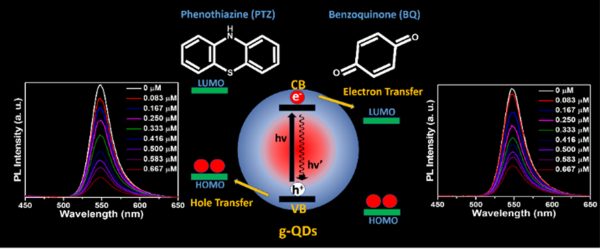 Dr Nimai Mishra, Assistant Professor, Department of Chemistry, SRM University-AP, Andhra Pradesh, along with his research group comprising of students pursuing PhD under him, Mr Rahul Singh, Mr Syed Akhil, and Ms V. G. Vasavi Dutt, have published a research article titled “Study of Shell Thickness Dependent Charge Transfer Dynamics in Green Emitting Core/Shell Giant Quantum Dots” Nature Index journal, “Inorganic Chemistry” published by The American Chemical Society having an impact factor of 5.1.
About the paper:
The superior photostability enables green-emitting graded alloy core/shell giant quantum dots (g-QDs) for optoelectronic application. However, it is essential to understand how the shell thickness affects interfacial charge separation. This work explores the impact of shell thickness on photoinduced electron transfer (PET) and photoinduced hole transfer (PHT) with an electron acceptor benzoquinone and a hole acceptor phenothiazine, respectively. The four graded alloy core/shell green-emitting g-QDs with different shell thicknesses were synthesised. The PET and PHT rate constants were obtained from photoluminescence and PL-lifetime decay measurement. Our study concludes that g-QDs with a diameter ~7.14 show a substantial improvement in charge transfer than g-QDs ≥ 8.5 nm in diameter. Similarly, the PET and PHT rates are 3.7 and 4.1 times higher for 7.14 nm g-QDs than for the 10.72 nm sample. The calculated electron and hole transfer rate constant (ket/ht) of g-QD with 7.14 nm in diameter are 10.80 × 10⁷ s-1 and 14 × 107 s-1, which shows 8.5 and 8 times higher compared to g-QDs with a 10.72 nm in diameter.
Industrial implications:
More importantly, these results highlight the impact of shell thickness on the excited state interactions of green-emitting g-QDs and conclude that g-QDs with a relatively thin shell can be a better choice as photoactive materials for photocatalysts, photodetectors, and solar cells.
Want the complete details of Dr Nimai Mishra’s paper?
Continue reading →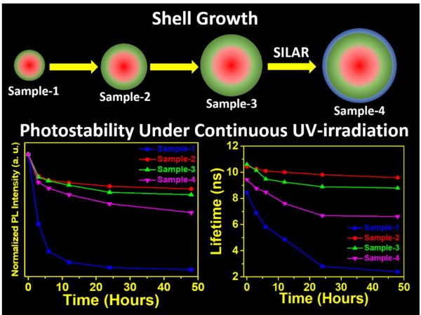 SRM University-AP is pleased to announce that Dr Nimai Mishra, Assistant Professor, Department of Chemistry, SRM University-AP, Andhra Pradesh, along with his research group comprising of students pursuing PhD under him, Mr Rahul Singh, Mr Syed Akhil, and Ms V.G.Vasavi Dutt, has published a research article titled “Shell thickness-dependent photostability studies of green-emitting “Giant” quantum dots” in the journal Nanoscale Advances (The Royal Society of Chemistry) with an impact factor of ~4.533.
About the research:
Highly efficient green-emitting core/shell giant quantum dots have been synthesized through a facile “one-pot” gradient alloy approach. Furthermore, an additional ZnS shell was grown using the “Successive Ionic Layer Adsorption and Reaction” (SILAR) method. Due to the faster reactivity of Cd and Se compared to an analogue of Zn and S precursors it is presumed that CdSe nuclei are initially formed as core and gradient alloy shells simultaneously encapsulate the core in an energy-gradient manner and eventually thick ZnS shells were formed. Using this gradient alloy approach, we have synthesized four different sized green-emitting giant core-shell quantum dots to study their shell thickness-dependent photostability under continuous UV irradiation, and temperature-dependent PL properties of nanocrystals. There was a minimum effect of the UV light exposure on the photostability after a certain thickness of the shell. The QDs diameter of ≥ 8.5 nm shows substantial improvement in photostability compared to QDs with a diameter ≤ 7.12 nm when continuously irradiated under the strong UV light (8 W/cm2, 365 nm) for 48 h. The effect of temperature on the photoluminescence intensities was studied with respect to shell thickness. There were no apparent changes in PL intensities observed for the QDs ≥ 8.5 nm, on the contrary, for example, QDs with < 8.5 nm in diameter (for ~7.12 nm) show a decrease in PL intensity at higher temperatures ̴90°C.
More importantly, these results highlight the synthesized green-emitting gradient alloy QDs with superior optical properties can be used for highly efficient green emitters and are potentially applicable for the fabrication of green LEDs.
Read the full paper: https://pubs.rsc.org/en/content/articlelanding/2021/NA/D1NA00663K
Continue reading →