An Algorithm that Redefines Employee Attrition Prediction Technology
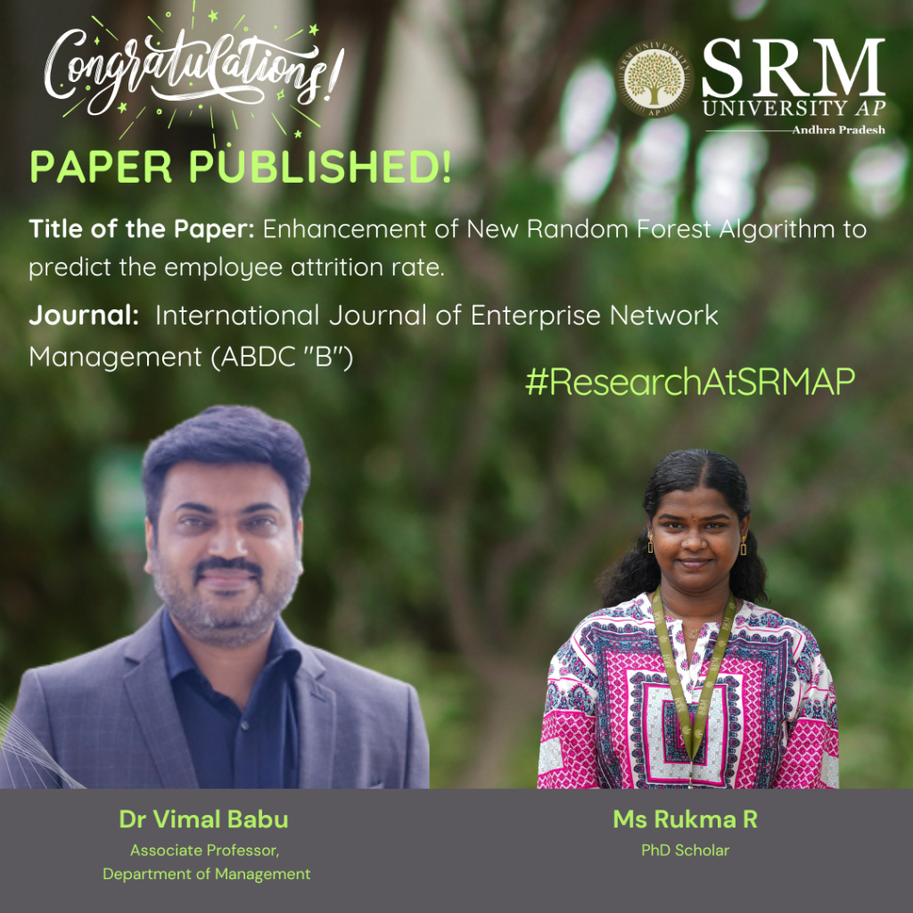
Dr Vimal Babu, Associate Professor in the Department of Management along with his PhD scholar Ms Rukma R, have co-authored a research paper titled “Enhancement of New Random Forest Algorithm to Predict the Employee Attrition Rate.” Published in the International Journal of Enterprise Network Management (ABDC-B), the paper also includes contributions from Dr Vijaya Prabhagar M, Assistant Professor at IIM Jammu.
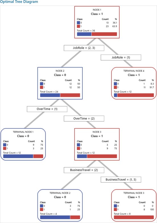
Their work focuses on improving machine learning techniques to better predict employee attrition, that is, the employees who are likely to leave the organisation. The study focuses on the impact of HR analytics adoption by applying algorithms to predict employee attrition. It uses the Random Forest algorithm to identify reasons for attrition. However, this algorithm can be slow due to the large number of decision trees it uses. To improve it, the study proposes a new method that selects the best decision trees using a technique called fractional factorial design, which outperforms all others in predicting employee attrition.
Abstract
The problem of employee attrition in every organisation concerns the employee turnover ratio, thereby increasing the cost of investment in human resources. Various factors are reasonable for the rapid attritions at different phases. The purpose of the current study is to predict the employees who are likely to leave the organization. The factors that lead to attrition are identified using the Random Forest algorithm. The Random Forest algorithm is a widely used supervised machine-learning technique for classification and prediction. However, the random forest algorithm has certain issue like it is too slow and ineffective for real time predictions. i.e., the large number of trees can make the algorithm, which results in a slower model. Therefore, the study proposes, a new alternative for choosing the appropriate decision trees based on the concept of fractional factorial design of experiments. The different performance criteria were compared across the modified random forest algorithm, existing random forest algorithm, Support Vector Machine (SVM), Logistic Regression (LR), Navie Bayes (NB), K – Nearest Neighbour (K-NN), Decision Tree, XG Boost tree and Neural Network (NN). It was found that the modified random forest algorithm excelled in all criteria and performed better than the existing ones.
Practical Implementation/Social Implications of the Research
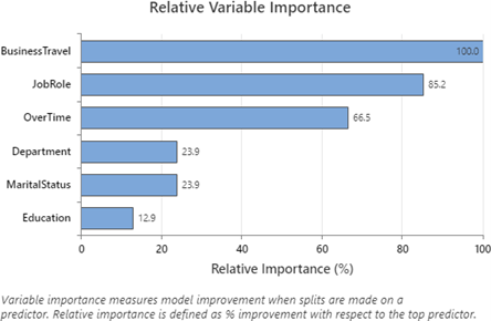
Practical implication: It emphasises the importance of focusing on factors like Business Travel, Job Role, Over Time, Department, Marital Status, and Education to reduce employee attrition. Organisations should design retention programs that support frequent travellers, manage overtime effectively, and address specific departmental needs. Tailoring job roles, promoting work-life balance through flexible hours, and providing educational opportunities are key strategies for enhancing employee satisfaction and retention.
Social Implication: The measures of this study can transform work culture into a more supportive and inclusive environment, promote gender equality by supporting employees with family responsibilities, and contribute to economic stability through reduced attrition. By investing in employee well-being, organizations can also strengthen their community engagement and corporate social responsibility initiatives for both employees and the broader society.
Future Research Plans
Future research could explore other factors influencing employee attrition, such as organizational culture, leadership style, and employee engagement. Longitudinal studies could track changes over time to understand the long-term effects on attrition rates. Additionally, investigating the interplay between identified factors, such as how Job Role and Over Time influence attrition, could provide deeper insights. Expanding the study across different industries and locations may reveal specific trends, allowing for tailored retention strategies. Lastly, incorporating qualitative methods like interviews and focus groups could offer a richer understanding of employees’ experiences and perceptions, complementing the quantitative findings.
- Published in Departmental News, News, Paari Current Happenings, Research News
Dr Amit Chakraborty and Scholar Publish their Research in Nature Index Journal
Dr Amit Chakraborty, Assistant Professor in the Department of Physics and Ms Shreecheta Chowdhury, PhD Scholar, have co-authored a research paper titled “Boosted top tagging through flavour-violating interactions at the LHC”, which has been published in the European Physical Journal C (Nature Index), Q1 Journal having an impact factor 4.2.
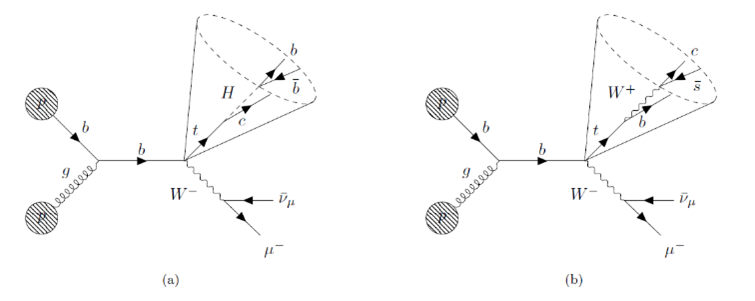
Their research focuses on identifying a rare process involving the top quark, one of the heaviest particles. In this rare decay, the top quark transforms into a charm quark and a Higgs boson, which then breaks down into two b quarks. The team uses the data collected at the Large Hadron Collider (LHC), the world’s largest particle smasher, and employs Machine Learning techniques to investigate the possibility of understanding these collision events and identifying signatures of physics Beyond the Standard Model.
Abstract
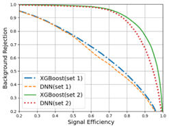
This paper describes a method for detecting a rare top quark decay into a charm quark and a Higgs boson (H), which decays further into b quarks, at the Large Hadron Collider (LHC), and introduces a tagging algorithm to identify boosted tops using large-R jets containing b- and c-tagged elements. We consider the associated production of the top quark with a W-boson and identify different observables to discriminate the signal from the Standard Model (SM) background events. Although our model with improved jet substructure methods outperforms existing approaches to tag such rare decay tops, the improvement in the New Physics reach in terms of t → cH branching ratio is marginal, even at the high luminosity run of LHC, compared to the existing limits from the LHC 13 TeV data. Additionally, the paper utilizes SHAP, a Game Theory-based method, to analyse the contribution of each observable to the classification of events, offering valuable insights into the classifier.
Future Research Plans
The team plans to explore Beyond Standard Model (BSM) physics through collider phenomenology. Leveraging the Higgs boson as a portal, they aim to investigate its potential role in dark matter interactions, neutrino mass generation, and possibly being part of a larger scalar sector. Additionally, Dr Chakraborty will delve into ultra-light particle searches and develop novel jet physics techniques using advanced Machine Learning (ML) algorithms for particle identification and classification. A crucial aspect of his work will be creating testable search strategies, ensuring direct comparison with experimental data from current and future colliders.
- Published in Departmental News, News, Physics News, Research News
SRM AP, Amaravati’s Landmark Collaboration with Carnegie Mellon University’s School of Computer Science, USA for AI Research, Education
SRM AP, Amaravati, is proud to announce a transformative five-year collaboration with Carnegie Mellon University’s School of Computer Science (CMU SCS), USA-one of the world’s foremost institutions in artificial intelligence (AI) and cutting-edge research. This strategic collaboration aims to push the boundaries of knowledge, innovation and education in AI-related disciplines, including machine learning, natural language processing, computer vision, infrastructure and systems, and AI ethics and policy.
At the heart of this collaboration is a shared vision to foster an ecosystem that nurtures groundbreaking research, cultivates exceptional talent, and accelerates advancements in AI-driven technologies.
A Pioneering Collaboration for AI Excellence

“CMU’s School of Computer Science is excited to work with SRM AP, Amaravati, on this landmark collaboration to advance research and bolster AI education. Together, we will shape the future of AI and empower the next generation of researchers, educators and industry leaders to push the frontiers of technology and drive meaningful change in society,” said Prof. Martial Hebert, Dean of CMU’s School of Computer Science.
Empowering Research Through Global Collaboration
As part of this collaboration, SRM AP, Amaravati’s research faculty and researchers will have the opportunity to engage directly with the esteemed faculty and researchers at CMU’s School of Computer Science. They will immerse themselves in CMU SCS’s pioneering AI labs, working alongside global experts in key research domains. This will facilitate research, knowledge sharing and the development of state-of-the-art AI innovations that address real- world challenges.

Dr P Sathyanarayanan, Pro-Chancellor of SRM AP, Amaravati, said that “To further strengthen research capabilities, this collaboration will also pave the way to establish advanced AI labs at SRM AP, Amaravati. These labs will be incubators for novel AI research, fostering a stimulating environment that promotes academic rigor, interdisciplinary collaboration and technological innovation”.
Advancing AI Education with World-Class Learning Opportunities
Beyond research, this collaboration is designed to enrich the academic experience of SRM AP’s teaching faculty and research scholars. Selected faculty members and scholars can audit cutting-edge AI courses at CMU’s School of Computer Science as visiting participants. This exposure will allow them to engage with CMU SCS faculty and contribute to developing robust AI curricula at SRM AP. They will also gain hands-on experience in designing assignments, worksheets and examinations that mirror real-world AI problem-solving scenarios, enhancing the quality of AI education at SRM AP, Amaravati,.
Unparalleled Research Internships for Students

Prof. Manoj K Arora, Vice Chancellor expressed, “In a move that underscores its commitment to nurturing future AI leaders, the collaboration will offer SRM AP students the opportunity to undertake research internships at CMU’s School of Computer Science.”
Selected students will spend approx. six weeks each summer immersed in a world-class research environment, gaining firsthand experience in tackling complex AI challenges alongside leaders in the field. This experience will provide students with unparalleled insights and exposure to global research methodologies, setting them apart in the highly competitive AI landscape.
By leveraging CMU SCS’s expertise and SRM AP’s commitment to academic excellence, this collaboration will drive innovation, expand knowledge horizons and create a lasting impact on the AI ecosystem between the universities.
Click here to know more about Institute >>
- Published in Collaborations, News, Research News
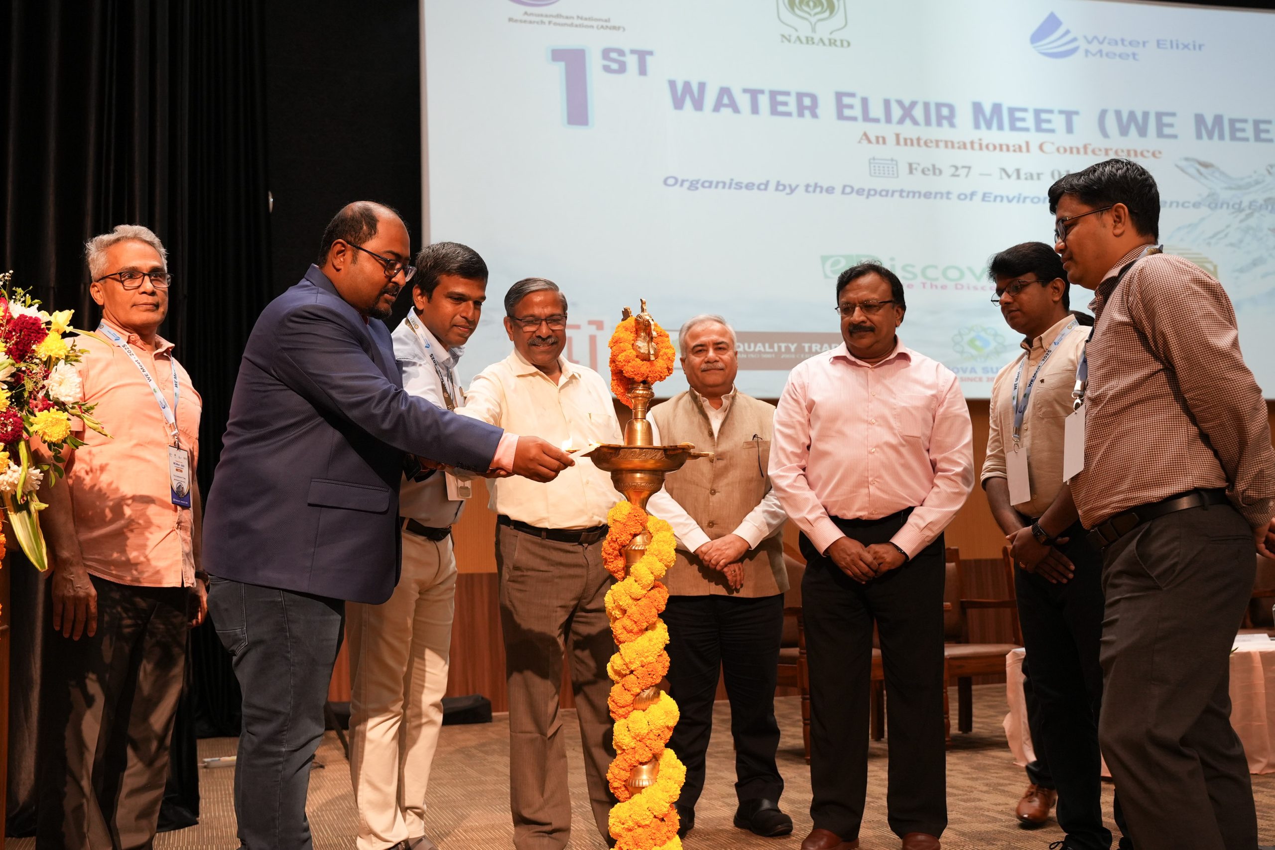
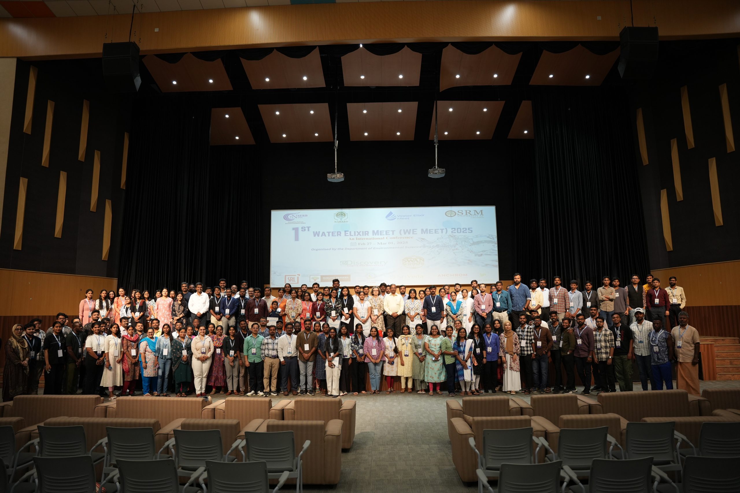
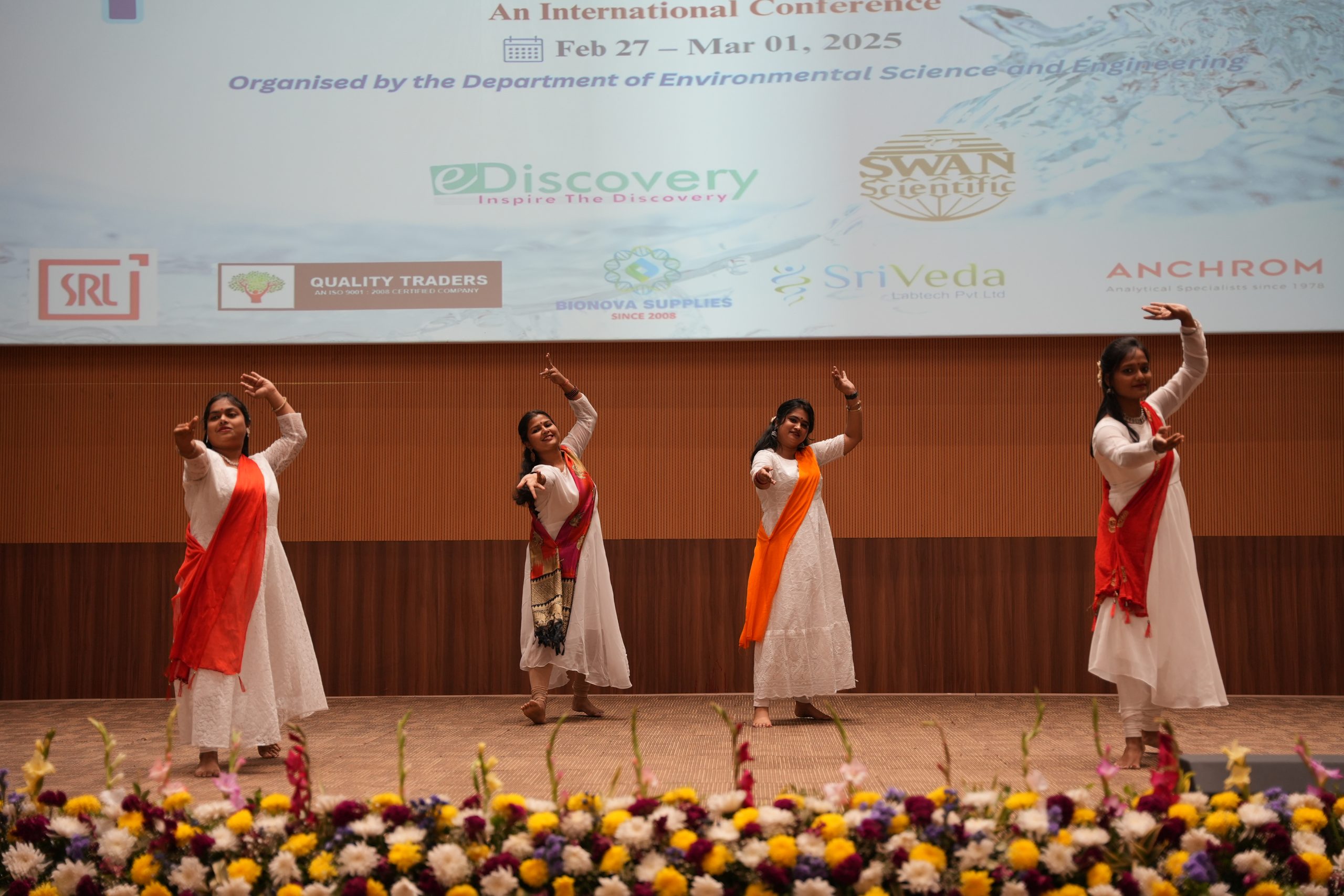
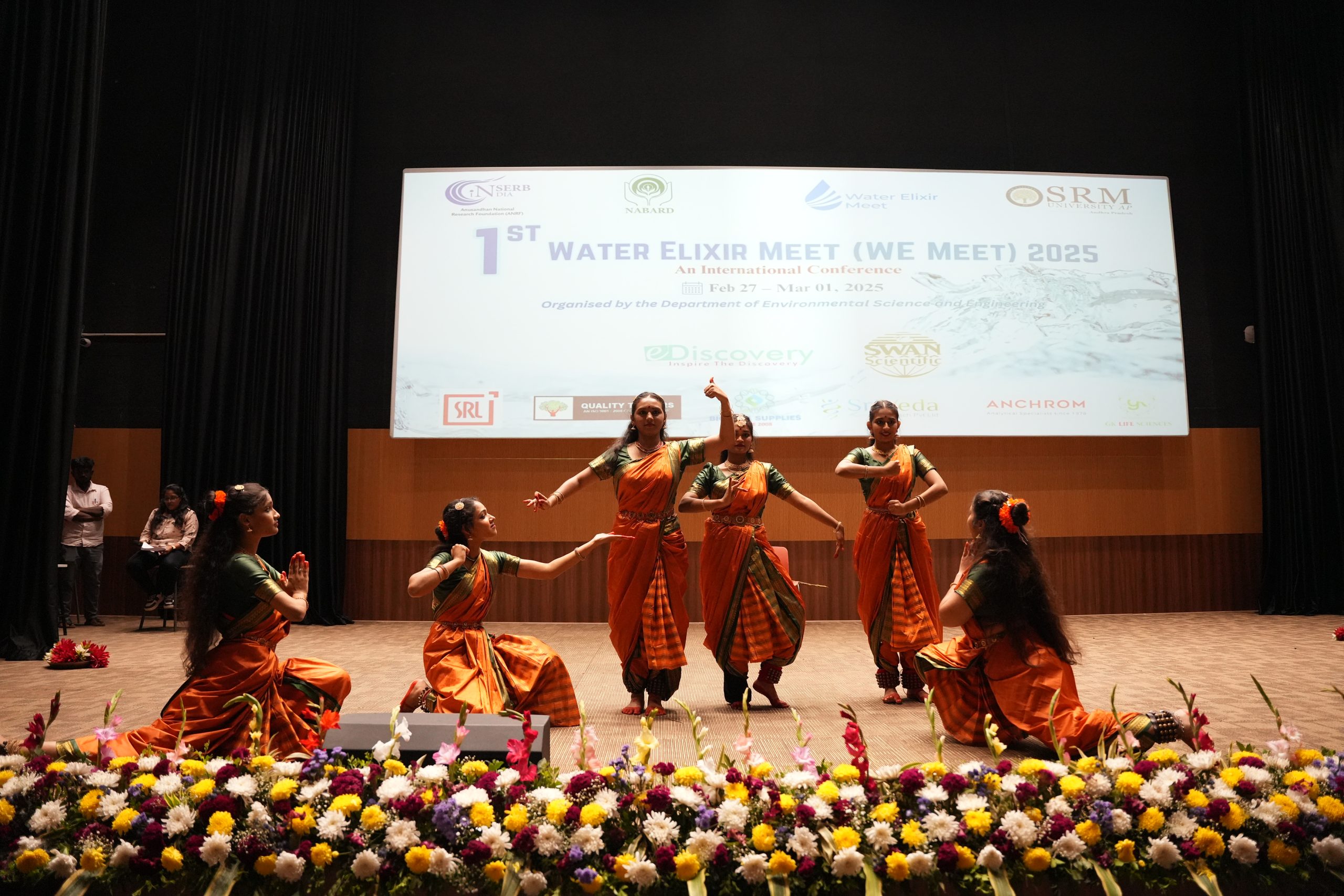
“A New Dawn of Water Sustainability”: 1st Water Elixir Meet 2025
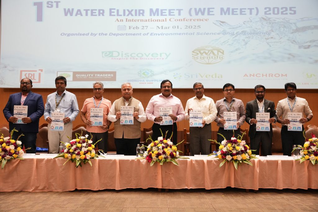
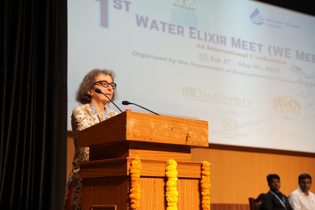
The Department of Environmental Science and Engineering hosted the 1st Water Elixir Meet (WE Meet 2025), a three-day international conference bringing together global minds to address one of the world’s most pressing challenges: water sustainability and security on Feb 27 – Mar 01, 2025. The conference was inaugurated by Prof. Rajasekhar Balasubramanian, Provost’s Chair Professor Group Head (Hydraulics, Hydrology and Climate Resilience), Department of Civil and Environmental Engineering, National University of Singapore.
“The available water quantity is decreasing, and the water quality is declining. There is a dire need to look into these parameters holistically and not separately. The We Meet 2025 is an ideal platform where scientists, researchers, policymakers, and industry leaders converge to address and manage global water resources efficiently and strategically,” stated Prof. Rajasekhar in his inaugural address.
WE Meet 2025 brought together more than 150 research abstracts and an esteemed lineup of global speakers, sharing groundbreaking insights into water resources, hydrogeology, and environmental sustainability. Prof. Kwang Ho-Choo from Kyunpook National University, South Korea, Prof. Shiao-Shing Chen from National Taipei University of Technology, Taiwan, Prof. Fulvia Chiampo from Politecnico di Torino, Italy, were some of the notable international speakers who delivered keynote sessions at the conference.
Prof. Manoj K Arora, Vice Chancellor, remarked beyond academic and research possibilities, WE Meet 2025 aimed at fostering global partnerships, innovative solutions, and cultural exchange. He stated, “WE Meet 2025 is a timely conference organised to address critical issues such as water resource management and water conservation.” Dr Rangabhashiyam Selvasembian, Head of the Department of Environmental Science and Engineering, also opined that this groundbreaking gathering fosters dialogue for impact. He said that the conference is a testament to the power of creative action in securing a sustainable tomorrow.
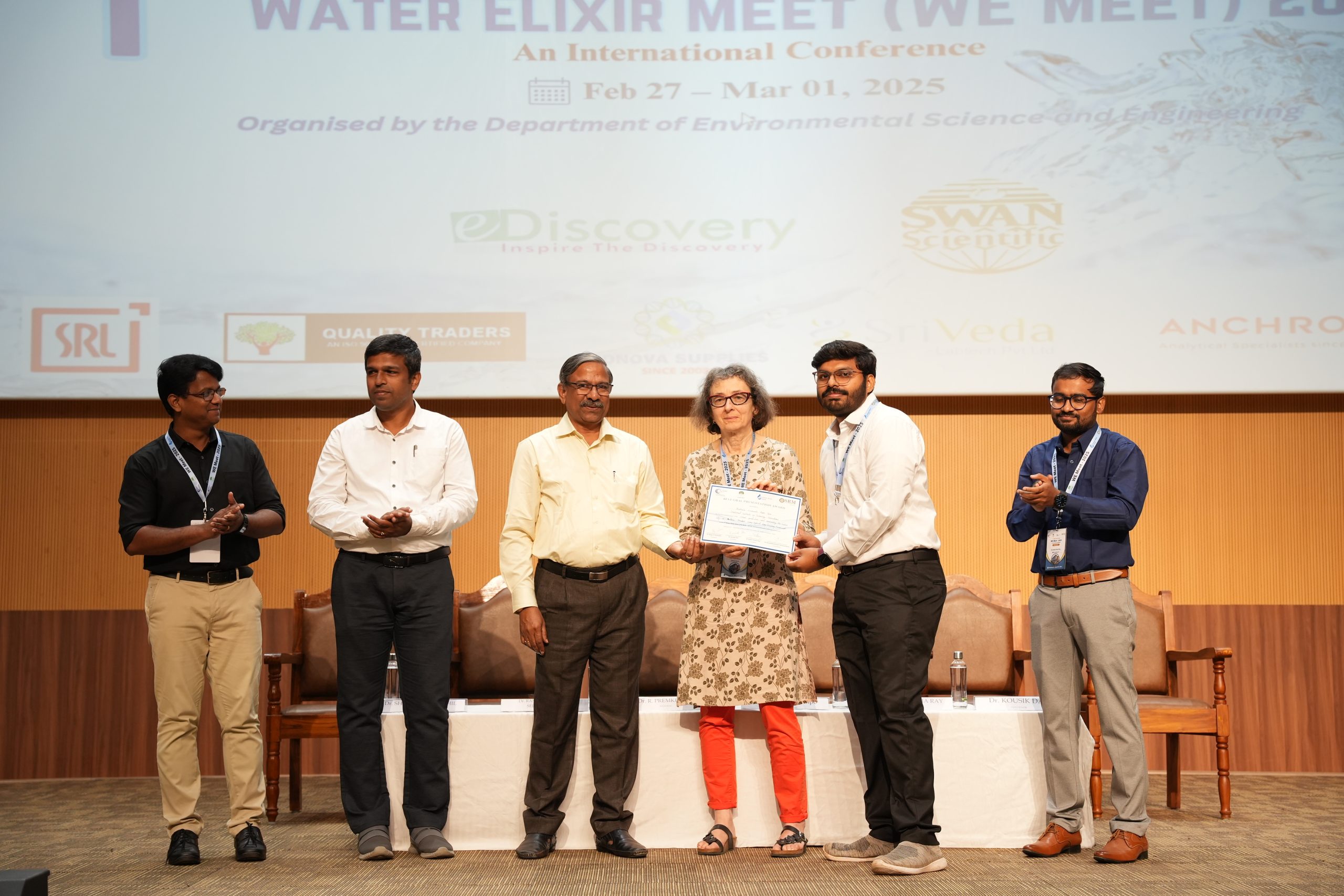
By showcasing cutting-edge technologies and driving policy advancements, WE Meet 2025 at SRM University-AP serves as a catalyst for real-world change. Exemplary research works such as the Best Oral Presentation and the Best Poster Presentation, were awarded top prizes at the valedictory ceremony. The conference also saw the participation of Registrar Dr R Premkunar, Dean – School of Engineering and Sciences, Prof. C V Tomy, Dean–Research, Prof. Ranjit Thapa, faculty, scholars and students of the varsity.
As the world grapples with increasing water challenges, this landmark international conference paves the way for a transformative journey to secure the future of water.
- Published in Departmental News, ENVS News, News, Research News
Trio Publish Patent on VR Based Therapy Systems
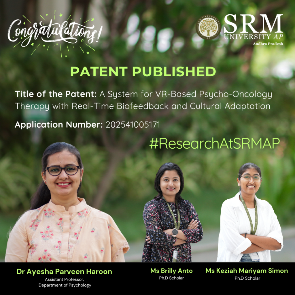 The Department of Psychology at SRM University-AP is proud to announce the publication of the patent titled, “A System for VR-Based Psycho-Oncology Therapy with Real-Time Biofeedback and Cultural Adaptation” by Dr Ayesha Parveen Haroon and her Research Scholars-Ms Brilly Anto and Ms Keziah Mariyam Simon. This groundbreaking research introduces a virtual reality therapy system that integrates real-time biofeedback.
Abstract
This research introduces a VR-based psycho-oncology therapy system that integrates real-time biofeedback and cultural adaptation to enhance the psychological well-being of cancer patients. The system utilizes virtual reality to create immersive therapeutic environments while continuously monitoring physiological indicators such as heart rate variability (HRV), skin conductance response (GSR), and respiratory rate to assess stress levels in real time. A processing unit analyzes this data using stress detection algorithms and selects personalized therapeutic interventions, including guided imagery, music therapy, mindfulness practices, and culturally relevant content. The system adapts dynamically to patient responses, ensuring an individualized approach to mental health support. Additionally, a cloud-based module securely stores therapy metrics for long-term monitoring and personalized care adjustments. This innovation offers a patient-centred, technology-driven intervention that enhances mental health outcomes, improves treatment adherence, and provides accessible psychological support for cancer patients in both clinical and home settings.
Explanation in layperson’s terms.
Cancer treatment can be emotionally challenging, leading to stress, anxiety, and depression. This research introduces a Virtual Reality (VR)-based therapy system that helps patients relax and manage stress through immersive environments like calming landscapes and guided meditation. The system monitors heart rate, breathing, and stress levels in real time and automatically adjusts therapy by playing soothing music, guided imagery, or relaxation exercises. It also includes culturally relevant content, making therapy more personalized and accessible, even for patients in remote areas. In short, this system acts as a virtual therapist, offering personalized mental health support to cancer patients during treatment and recovery.
Practical Implementation & Social Impact
Practical Implementation
Used in hospitals, cancer centers, and home-based care for stress management.
Helps psycho-oncologists and mental health professionals provide personalized therapy.
Supports palliative care and extends telehealth access to remote areas.
Social Implications
• Reduces stress and anxiety in cancer patients, improving their quality of life.
• Increases accessibility to mental health support, especially in underserved areas.
• Enhances treatment adherence by promoting emotional well-being.
• Offers culturally relevant therapy, making interventions more relatable and effective.
This system bridges the gap in psycho-oncology care, making mental health support more engaging, accessible, and personalized.
Future Research Plans
• Enhancing VR therapy with AI-driven personalization.
• Integrating EEG-based biofeedback for stress monitoring.
• Developing affordable, portable VR solutions for remote care.
• Conducting clinical trials for validation.
• Expanding telehealth access for home-based psycho-oncology care.
- Published in Departmental News, News, Psychology News, Research News
Parameters to Measure a University Entrepreneurial Ecosystem
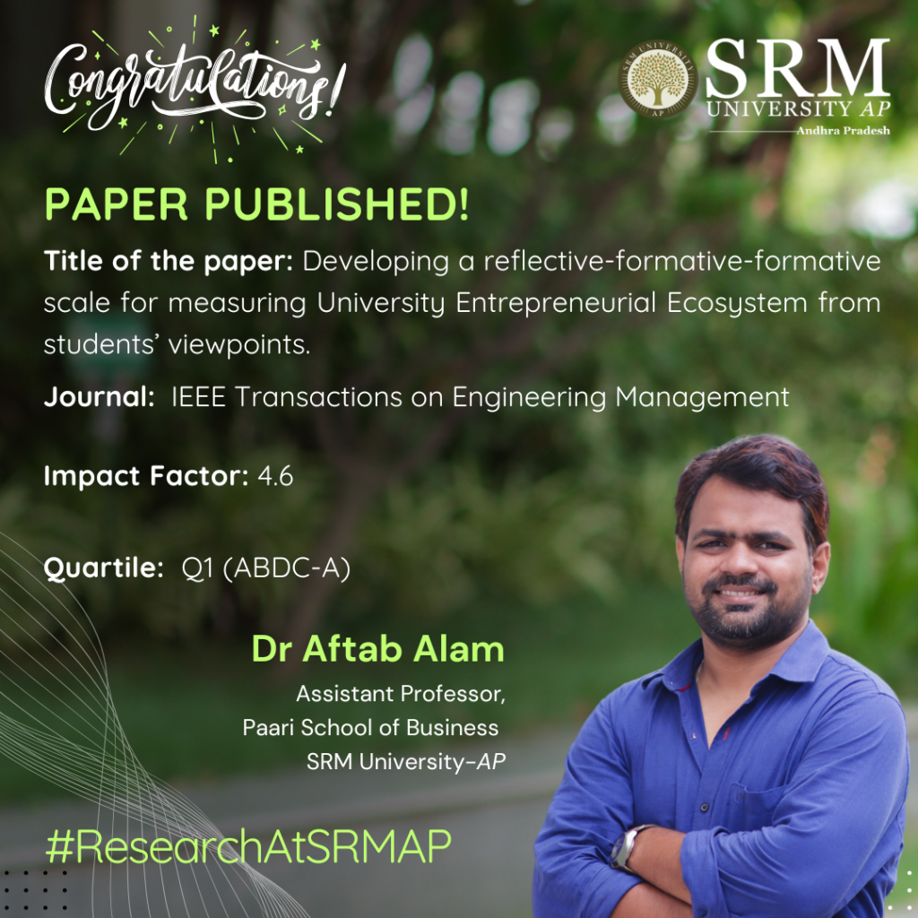
With the increasing emphasis on startup culture and enhancing entrepreneurial ventures among the youth, Dr Aftab Alam, Assistant Professor from the Department of Management has published a paper “Developing a Reflective-formative-formative Scale for Measuring University Entrepreneurial Ecosystem from Students’ Viewpoints” in the Q1 journal IEEE Transactions on Engineering Management having an impact factor of 4.6.
Dr Aftab and his team have conducted research on how universities can create the best environment to support entrepreneurship among students and staff. The team has identified three key elements that help an entrepreneurial ecosystem thrive:
- Skill Development – Helping people learn how to start and manage businesses.
- Resources – Providing tools, funding, and guidance.
- Culture – Encouraging creativity, risk-taking, and innovation.
They have also created a way to measure how well universities are doing in building this supportive environment, helping them improve and compare with others.
Abstract
This research addresses the gap in the literature on University Entrepreneurial Ecosystems (UEE) by conceptualizing UEE as an ecology-inspired system with dimensions like Entrepreneurial Skill Development, Resources, and Culture. Unlike prior studies focusing on isolated aspects, we provide a comprehensive framework and develop robust measurement scales using a rigorous four-step methodology, ensuring nomological validity. The paper contributes to entrepreneurial ecosystem literature by offering a novel conceptualisation and practical tool for evaluating and comparing UEE performance. This study aids scholars in understanding UEE’s holistic impact and supports managers in enhancing university-driven entrepreneurship for regional development.
Practical Implication of the Research
This paper contributes to the entrepreneurial ecosystem literature by conceptualising and measuring performance of university entrepreneurial ecosystems (UEE). Beyond academic circles, it could also serve as a valuable tool for managers seeking to evaluate and enhance the performance of these ecosystems.
Collaboration
- Dr Arpita Ghatak, Kent Business School, University of Kent
- Dr Bhaskar Bhowmick, Indian Institute of Technology Kharagpur
- Dr Swagato Chatterjee, Queen Mary University of London
Future Research Plan
- Longitudinal Validation: Conduct longitudinal studies to evaluate how UEE dimensions evolve over time and their sustained impact on entrepreneurial outcomes like spinoffs, startups, and regional development.
- Comparative Analysis: Compare UEEs across diverse geographic, cultural, and institutional contexts to identify patterns, best practices, and challenges unique to different ecosystems.
- Entrepreneurial Outcomes Measurement: Develop advanced metrics to assess the direct and indirect outcomes of UEEs on entrepreneurial intentions, skill-building, and economic growth.
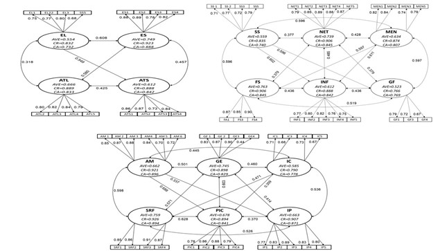
Confirmatory first-order measurement model results for the subscales
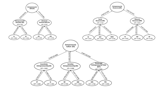
Hierarchical Third-Order Measurement Model Results
- Published in Departmental News, News, Paari Current Happenings, Research News
Revolutionising Cardiac Diagnostics and Real-time Health Monitoring
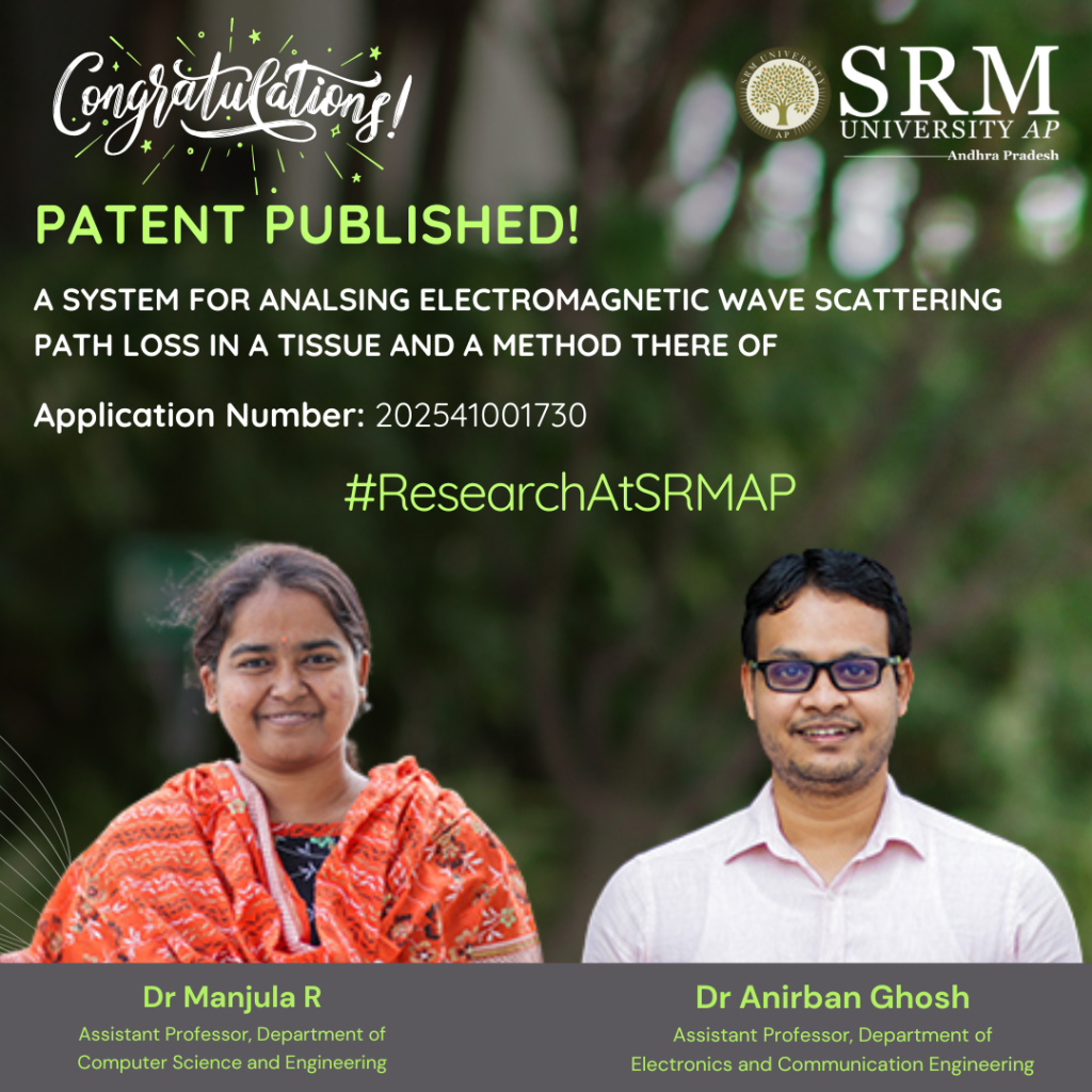
Dr Manjula R, Assistant Professor, Department of Computer Science and Engineering, and Dr Anirbhan Ghosh, Assistant Professor, Department of Electronics and Communication Engineering, has recently had their patent published titled “A System for Analyzing Electromagnetic Wave Scattering Path Loss in a Tissue and a Method Thereof” with Application no: 202541001730.
The faculty duo has revolutionised cardiac diagnostics and real-time health monitoring through their invention. This innovative system analyses electromagnetic wave scattering in biological tissues, using terahertz (THz) frequencies to optimise nanosensor communication and path loss analysis. By leveraging cutting-edge technology, it enables advanced biomedical devices for precise physiological monitoring and safer, more reliable in-vivo communication systems. A step forward for heart health and medical breakthroughs, this invention bridges the gap between technology and life-saving healthcare solutions.
- Published in CSE NEWS, Departmental News, ECE NEWS, News, Research News
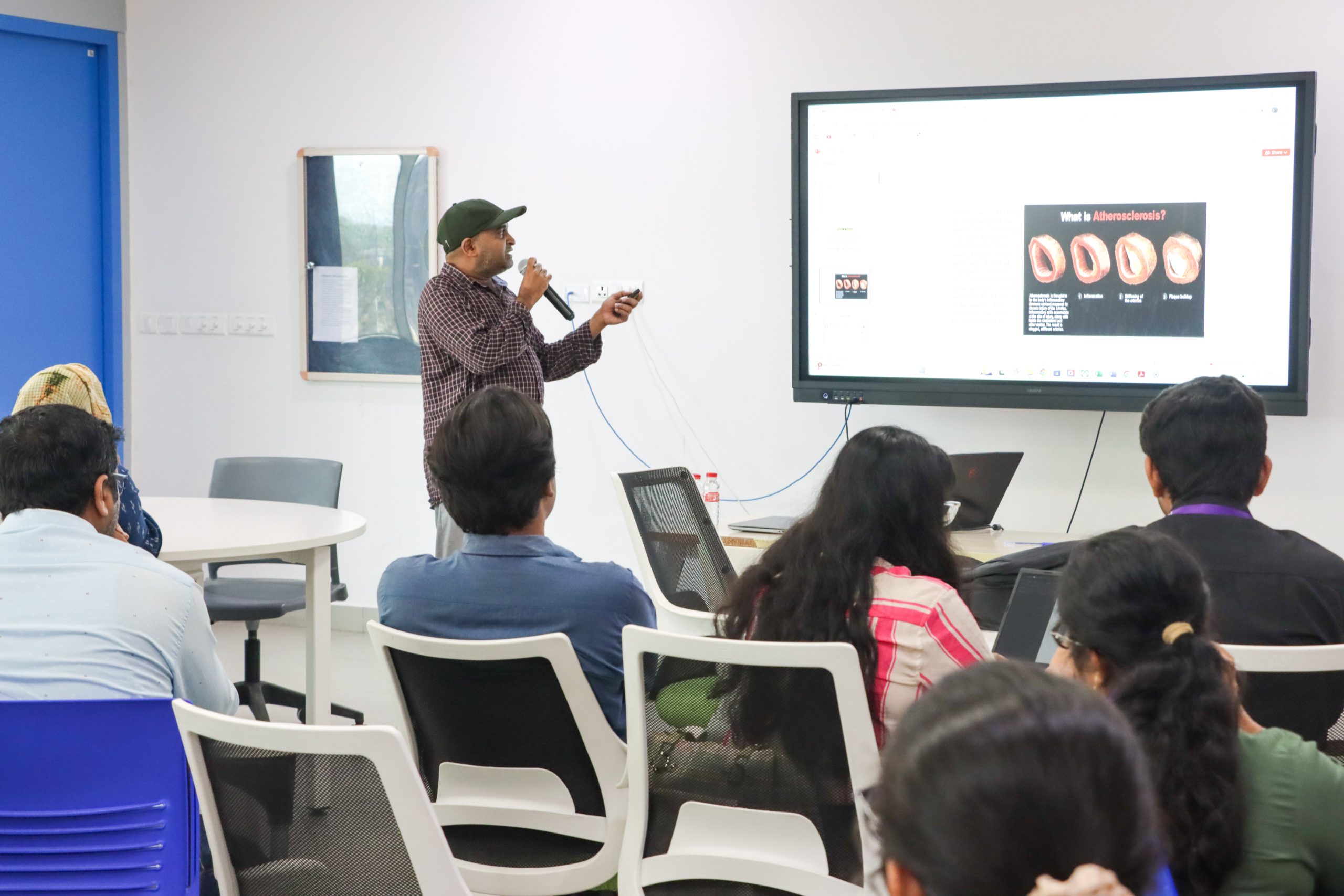
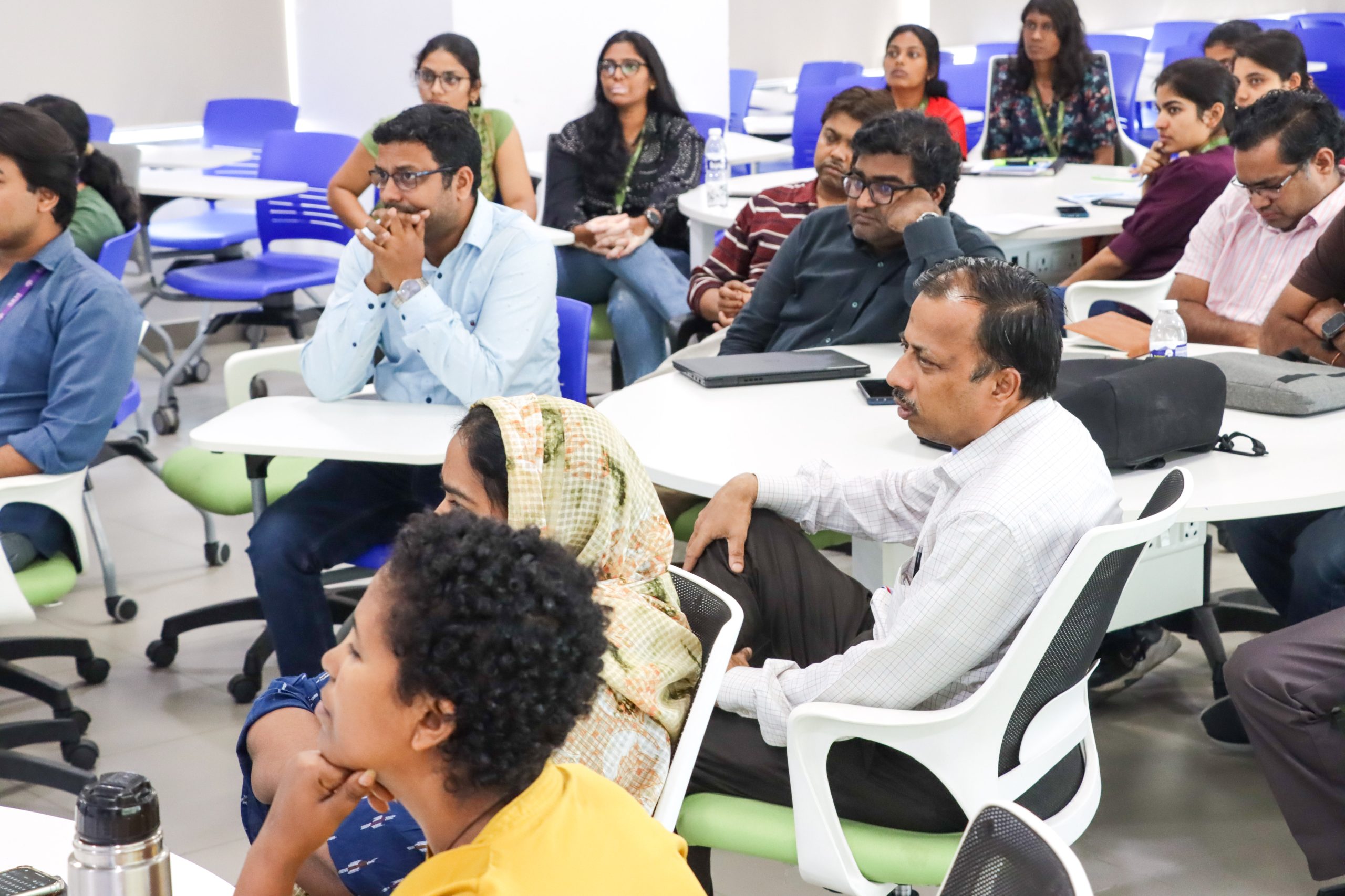
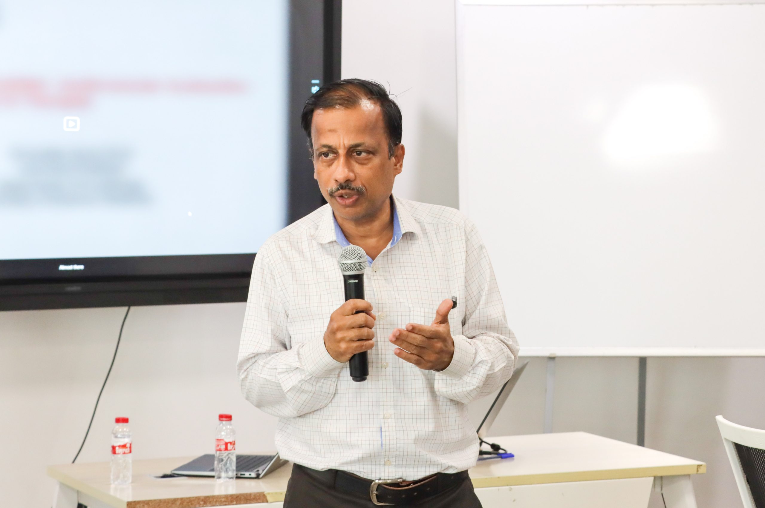
From Mechanisms to Theory: Dr Siva Reddy Addresses Students of Biological Sciences
 The Department of Biological Sciences organised a significant expert talk titled “Senescence-Associated Cardiovascular Dysfunction: From Mechanisms to Therapies”, the talk was led by Dr Kotla Siva Reddy, Associate Professor, Department of Cardiology, Division of Internal Medicine, The University of Texas MD Anderson Cancer Center, Houston, TX, USA. This talk aimed at research scholars in the field explored the complex interplay between cellular senescence, cardiovascular health, and the adverse effects associated with cancer and HIV treatment drugs.
The resource person, Dr Siva Reddy, with his extensive academic background in Biotechnology set the stage for discussions. Key topics of discussion included the molecular mechanisms underlying cardiovascular dysfunction linked to cancer and HIV therapies, particularly focusing on oxidative stress and endothelial dysfunction.
The event also fostered an interactive environment where scholars and students engaged in meaningful dialogues. Attendees gained crucial insights into how cellular senescence and foam cell formation contribute to atherosclerosis, thereby affecting cardiovascular health.
In conclusion, the event successfully facilitated rich discussions on the intersections of oncology, virology, and cardiovascular health. By focusing on innovative therapeutic strategies and the underlying mechanisms of cardiovascular diseases induced by treatment regimens, the event contributed significantly to enhancing the collective understanding and research efforts in the realm of cardiovascular aging.
- Published in Biology News, Departmental News, News, Research News
Dr Abhijit Dasgupta Publish his Research on Alzheimer’s disease (AD) in Nature Index Journal
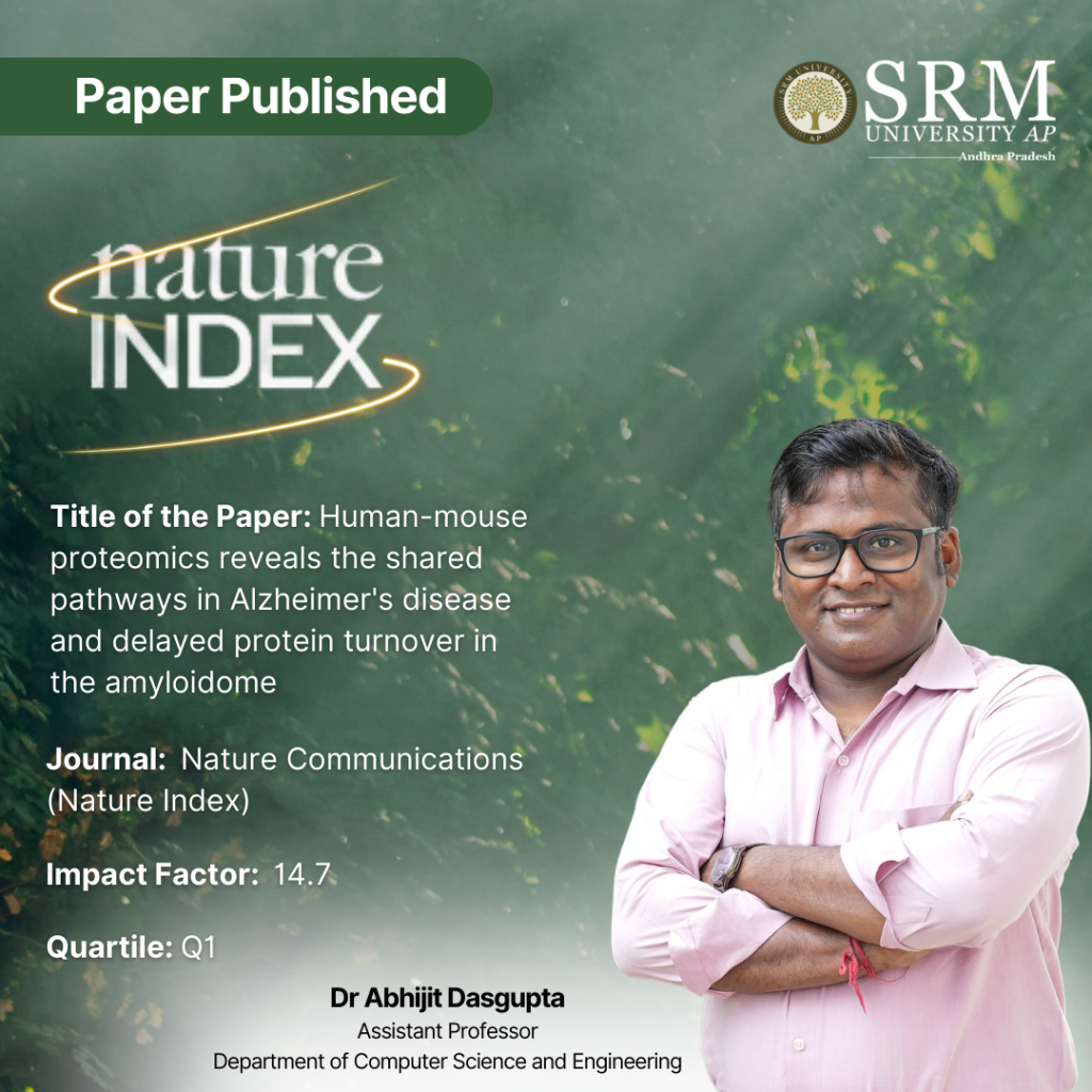
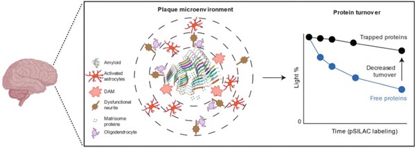
Dr Abhijit Dasgupta, Assistant Professor from the Department of Computer Science and Engineering, has published his groundbreaking research on using deep proteomics to analyse Alzheimer’s disease (AD) models in mice and comparison with human AD protein alterations. He has published the paper titled “Human-mouse proteomics reveals the shared pathways in Alzheimer’s disease and delayed protein turnover in the amyloidome” as one of the first authors in the Nature Index journal Nature Communications, having an impact factor of 14.7.
Abstract
Alzheimer’s disease (AD) is a complex neurodegenerative disorder characterized by amyloid-beta (Aβ) plaques and tau tangles. This study presents a comprehensive proteomic and phosphoproteomic analysis of multiple AD mouse models, comparing their molecular pathways with human AD data to evaluate their translational relevance. Using deep proteomics, the study identifies shared and distinct protein alterations across amyloidosis models (5xFAD, APP-KI), tauopathy models (3xTG), and splicing dysfunction models (BiG). While these models collectively replicate 42% of human AD protein alterations, amyloid formation significantly delays protein turnover in the amyloidome, contributing to proteome-transcriptome discrepancies. Proteomic and turnover analysis highlight the accumulation of proteins such as ApoE, CLU, and HTRA1, implicating lysosomal and autophagic dysfunction. These findings underscore the importance of protein homeostasis in AD pathology and provide a multi-omics resource for selecting appropriate mouse models for specific disease mechanisms.
Explanation of the Research in Layperson’s Terms
Alzheimer’s disease (AD) is a brain disorder that affects memory and thinking ability. Scientists know that AD is caused by harmful clumps of proteins—called amyloid-beta (Aβ) plaques—that build up in the brain. However, studying AD in humans is challenging, so researchers often use mice with genetic modifications that mimic the disease.
This study looks at different types of AD mouse models and compares their brain protein changes to those seen in humans with Alzheimer’s. The researchers analysed thousands of proteins to understand how the disease progresses, how mouse models reflect human AD, and what biological processes might be involved.
One key finding is that in both mice and humans, the buildup of Aβ plaques slows down the normal breakdown and recycling of certain proteins. This means that some proteins accumulate in the brain not just because they are being produced in excess but also because they are not being cleared efficiently. This could explain why some aspects of AD develop over time.
The study also shows that while current mouse models capture some aspects of human AD, none of them fully replicate the disease. However, when different models are combined, they represent a larger portion of the changes seen in human AD. This research provides valuable insights into how AD develops and helps scientists choose the right mouse models for studying different parts of the disease. It also highlights potential targets for future treatments focused on restoring protein balance in the brain.
Practical Implementation/Social Implications of the Research
Practical Implementation
This research helps improve Alzheimer’s disease (AD) drug development by identifying key proteins involved in disease progression. It aids in selecting better animal models for testing treatments, enhances early diagnosis through biomarkers, and supports AI-driven modelling of disease progression. The findings could lead to therapies that improve protein clearance, slowing AD progression.
Social Implications
With AD cases rising globally, this study has significant public health and economic impacts. It could reduce caregiver burden, lower healthcare costs, and improve the quality of life for ageing populations. Additionally, it encourages ethical advancements in research by promoting better human-relevant models and minimising reliance on animal testing.
Collaborations
- St. Jude Children’s Research Hospital, Memphis, TN, USA.
- University of Tennessee Health Science Center, Memphis, TN, USA
- Yale University School of Medicine, New Haven, CT, USA
Future Research Plans
The future focus will be on AI-driven modelling of temporal proteomics to understand Alzheimer’s Disease (AD) progression and identify therapeutic targets. By integrating mass spectrometry-based proteomics, machine learning, and protein turnover analysis, the goal is to bridge transcriptome-proteome discrepancies and uncover key regulatory pathways.
The research aims to develop cost-effective AI-based diagnostics for AD and cardiomyopathies, utilizing multi-omics integration for early detection and personalized treatments. This approach will contribute to precision medicine and scalable healthcare solutions for a broader impact.
- Published in CSE NEWS, Departmental News, News, Research News
Patent on Pneumonia Diagnosis Using Deep Learning Techniques
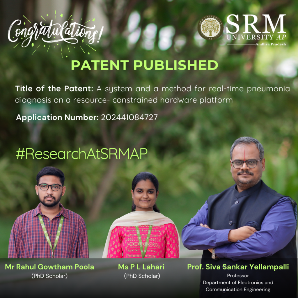 In the patent titled “A System and a Method For Real-Time Pneumonia Diagnosis On a Resource- Constrained Hardware Platform,” authored by Prof. Siva Sankar Yellampalli from the Department of ECE and his research scholars – Mr Rahul Gowtham Poola and P L Lahari, a novel diagnostic solution is presented to enhance pneumonia detection in low-resource settings. With Application No: 202441084727, this research explores the integration of advanced deep learning techniques with a compact microcontroller-based system, providing an innovative approach to improve healthcare accessibility and prompt medical intervention.
Abstract:
The research focuses on the development of an innovative system for real-time pneumonia diagnosis leveraging advanced deep learning techniques integrated with edge computing technology. The proposed solution employs the MAX78000 microcontroller, a resource-constrained hardware platform, to deploy a sophisticated neural network model capable of analyzing chest X-ray images. The invention addresses the pressing need for accessible, cost-effective, and efficient diagnostic tools in under-resourced and remote environments. The system encompasses a complete diagnostic pipeline, including image acquisition via an onboard parallel camera module, real-time image processing, and display of results on a 3.5″ touch-enabled TFT screen. The deep learning model, optimized for the constraints of the MAX78000, performs real-time classification of chest X-ray images into either normal or pneumonia-affected categories. By operating entirely on-device, the system eliminates the need for high-power servers or internet connectivity, thereby reducing latency and dependency on external infrastructure. This research emphasizes portability, energy efficiency, and low-cost deployment, making the solution highly suitable for primary healthcare facilities, rural clinics, mobile health units, and disaster-response scenarios. With the ability to deliver immediate, accurate diagnoses, the device significantly enhances clinical decision-making and enables timely medical intervention. Additionally, the scalable and adaptable design of the system opens possibilities for broader medical imaging applications, extending its utility beyond pneumonia diagnostics. Experimental results showcase the performance of the neural network model, demonstrating prediction accuracies ranging between 66% and 97% for different test cases on the MAX78000 microcontroller. These findings underline the potential of the proposed system as a transformative tool for advancing point-of-care diagnostics in low-resource settings.
Explanation in Layperson’s terms.
The research presents a compact, affordable device that helps doctors quickly detect pneumonia by analyzing chest X-ray images in real-time. It uses advanced artificial intelligence (AI) technology, called deep learning, to examine the X-rays and determine whether a patient has pneumonia or not. What makes this device special is that it works entirely on a small, low-power microcontroller called the MAX78000, instead of needing powerful computers or internet access. The process begins when the device captures a chest X-ray image using its built-in camera. Then, the AI model, which has been trained to recognize patterns associated with pneumonia, analyzes the image. The results are displayed instantly on a small screen, allowing healthcare providers to make quick decisions. This real-time diagnosis can be life-saving, especially in emergency or rural settings where access to advanced medical equipment or high-speed internet is limited. Technically, this system combines AI and edge computing, meaning all the heavy processing happens directly on the device rather than in remote servers. This design keeps costs low, ensures patient data privacy, and makes the device highly portable and energy-efficient. The technology can work even in places with unreliable electricity, making it ideal for use in mobile health units, rural clinics, or disaster zones. Additionally, the invention can be adapted for diagnosing other diseases, showcasing its versatility in improving healthcare globally.
Practical Implementation
This research can be practically implemented as a compact, standalone device for diagnosing pneumonia in healthcare settings where access to advanced medical equipment is limited. It works as follows:
- Deployment in Rural Clinics and Mobile Health Units: The device can be used in clinics in remote or underserved areas where large X-ray machines and advanced computing resources are unavailable. It provides on-the-spot diagnosis.
- Point-of-Care Diagnostics: The portability and integration of image acquisition, AI-based processing, and display into a single unit make it ideal for bedside use in hospitals or during emergency care.
- Disaster Response: Its low-power and internet-free design make it a critical tool in disaster zones, refugee camps, or any setting where power and connectivity are unreliable.
- Telemedicine Integration: The device can complement telemedicine by providing accurate diagnostic results to remote doctors, helping bridge the gap between frontline healthcare workers and specialists.
Social Implications
Improved Access to Healthcare: By making pneumonia diagnosis accessible in rural and underserved regions, this device can drastically reduce the gap in healthcare services between urban and remote areas. It empowers healthcare providers in low-resource settings to deliver timely diagnoses.
- Affordability: The use of a low-cost microcontroller ensures that the device is affordable for governments and healthcare organizations, particularly in developing countries. This can enhance healthcare access for low-income populations.
- Reduced Mortality Rates: Pneumonia is a leading cause of death in children under five and elderly individuals, especially in low-income countries. This device’s ability to provide real-time, accurate diagnosis allows for earlier intervention and treatment, potentially saving countless lives.
- Privacy and Security: Since all data is processed locally on the device, it ensures patient privacy by eliminating the need to transfer sensitive medical data to cloud servers, addressing concerns about data security.
- Scalability: The underlying technology can be adapted to diagnose other diseases, creating a broader impact on global health. For example, similar systems could be used for tuberculosis, COVID-19, or other respiratory conditions, further enhancing healthcare infrastructure.
By addressing critical gaps in diagnostic capabilities and ensuring accessibility and affordability, this research has the potential to transform healthcare delivery and improve quality of life, especially in marginalized communities.
Collaborations:
Rahul Gowtham Poola, Ph.D Scholar, Dept of ECE, SRM University-AP
P.L. Lahari, Ph.D Scholar, Dept of ECE, SRM University-AP
Prof. Siva Sankar Yellampalli, Professor of Practice, Dept of ECE, SRM University-AP
- Published in Departmental News, ECE NEWS, News, Research News